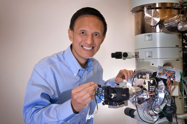
The 2024 MSC-SMC meeting will be featuring leading scientists in the fields of light and electron microscopy and their applications to biology and material science. Their expertise in these topics represents some of the most up to date advancements and applications in routinely used microscopy techniques.

Plenary Speaker

Let's celebrate our 50th anniversary with inspiring researchers passionate about Microscopy.

  • Dr. James Weaver

    Dr. James Weaver

    MIT & Harvard University

    Dr. James C. Weaver runs the Wide-Field Electron Optics Laboratory and leads the Biologically Inspired Materials and Design Group. He received his Bachelor’s degree in Aquatic Biology and Ph.D. in Marine Science from the University of California, Santa Barbara, and went on to pursue postdoctoral studies in Molecular Biology, Chemical Engineering, Physics, and Earth History. Working at the interface between zoology, materials science, biomedical engineering, and multi-material 3D printing, his main research interests focus on investigating structure-function relationships in hierarchically ordered biological composites and the advanced fabrication of their synthetic analogues. He has played critical roles in the development of new model systems for the study of a wide range of biomineralization processes, is an internationally recognized and award-winning scanning electron microscopist, and for ten years has led additive manufacturing research efforts on commercial 3D printers within the greater Harvard community. With a strong history of national and international academic and industrial collaborations, he has coauthored more than 150 journal articles in the biological, physical, and geological sciences. His work has been featured on the covers of more than 40 scientific journals and he has contributed to numerous collaborative art installations, which have been exhibited in Berlin, Boston, Frankfurt, London, New York, Paris, and San Francisco.

  • Dr. Nathalie Nguyen-Quoc Ouellette

    Dr. Nathalie Nguyen-Quoc Ouellette

    Université de Montréal

    Nathalie Nguyen-Quoc Ouellette is an astrophysicist, science communicator and lifetime lover of all things space! She obtained her Ph.D. in Physics & Astronomy at Queen's University in Kingston, Ontario in 2016. Her research focuses on galaxy formation and evolution, particularly those found in clusters. Nathalie is currently the Deputy Director of the Trottier Institute for Research on Exoplanets (iREx) and the Mont-Mégantic Observatory (OMM) at the University of Montréal and is also the Outreach Scientist for the James Webb Space Telescope in Canada collaborating with the Canadian Space Agency. She is a frequent contributor and analyst in Canadian media on everything related to space. She also organises and participates in science outreach events from local to international scales to encourage the interest and participation of youth and the general public in space science and to increase scientific literacy in Canada.

    Dr Ouellete will be our keynote speaker during the Banquet at the Canada Science and Technology Museum. This will be the first milestone between our communities in the hope of initiating exchanges that could lead to innovation in our respective fields.

  • Dr. Anja Geitmann

    Dr. Anja Geitmann

    Department of Plant Science - McGill University

    Professor in the Department of Plant Science - McGill University. She holds the Canada Research Chair in Biomechanics of Plant Development and she uses high-end light and electron-based imaging for the investigation of plant architectural features at multiple scales. Her team combines structural data with micromechanical testing and finite element modeling to interrogate how cell morphogenetic processes lead to the formation of functional tissues. Dr. Geitmann served as the President of the MSC from 2013-2015.

    To celebrate the 50th anniversary of the MSC, Dr. Anja Geitmann will highlight key moments in the history of microscopy in Canada and walk down memory lane illustrating how dedication and enthusiasm led to the building of a community of researchers with highly diverse goals but a common passion

  • Dr. John Rubinstein

    Dr. John Rubinstein

    The Hospital for Sick Children in Toronto

    A native of Toronto, John Rubinstein received his BSc from the University of Guelph, graduating in 1998. For his PhD he moved to England to work with Dr. Richard Henderson and Sir John Walker at the MRC laboratories in Cambridge. He completed his PhD research and a postdoctoral fellowship before returning to Canada for a second short postdoctoral fellowship at U of T. He started his own research group at The Hospital for Sick Children in 2006, where he holds a Canada Research Chair, and studies the structural biology of bioenergetics and develops new methods in cryoEM to facilitate this work. He has been recognized by the Burton Medal of the Microscopy Society of America, the New Investigator award of the Canadian Society of Molecular Biosciences, the Lars Ernster Lectureship in Bioenergetics, and most recently a Doctorate of Philosophy honoris causa from Stockholm University.

    Talk Summary: Single particle electron cryomicroscopy (cryo-EM) has evolved from a technique capable only of low-resolution insight into molecular structure into what is now often the most straightforward method for determining high-resolution structures of macromolecules and macromolecular assemblies. Canadian scientists have made important contributions to this field from its beginning, and there are now many laboratories around the country using this technology. In this talk I will outline the growth of cryo-EM in Canada. I will discuss the state-of-the-art and some new approaches using the cryo-EM with examples from my own laboratory.

  • Jim Corbett

    Jim Corbett

    University of Waterloo

    Jim Corbett is a Professor Emeritus from the Department of Physics and Astronomy in the University of Waterloo. Over thirty years, with colleagues at Waterloo, he studied the structures of a variety of crystalline materials by transmission electron microscopy, concluding with an investigation of charge density waves in transition metal chalcogenides. Dr Corbett served as president of the MSC in the period 1995-1997.

    To celebrate 50 years of our MSC/SMC, Dr Corbett will offer reminiscences of the early days of electron microscopy.

Parallel Sessions - Biology

  • Dr. Elitza Tocheva

    Dr. Elitza Tocheva

    Department of Microbiology and Immunology - University of British Columbia

    Research in my lab combines advanced structural approaches with in-depth molecular, biochemical and bioinformatic analyses to elucidate the role and function of essential microbial components and macromolecular assemblies. We develop and implement approaches such as correlative light and electron microscopy, super resolution light microscopy and cryo-electron tomography.

    Talk Title: Advanced imaging approaches to characterize microbial ultrastructure

  • Dr. Joaquin Ortega

    Dr. Joaquin Ortega

    Department of Anatomy and Cell Biology - McGill University

    Joaquin Ortega received his PhD in Biochemistry and Molecular Biology in 1999 for his studies in the ultrastructure of influenza virus ribonucleoproteins. Joaquin joined the Faculty of Health Sciences at McMaster in the Department of Biochemistry and Biomedical Sciences on July 2003. In 2017, Joaquin moved his laboratory to McGill. Today, in addition to working with his research team, he is also the Research Director of the Facility for Electron Microscopy Research (FEMR).

    Talk Summary: Regulatory mechanisms in specialized ribosomes in neurons. Ribosomes are cellular machines that translate the genetic code to make proteins in all types of cells. In neurons, this process needs special regulation so that proteins are made at the synapse, a critical connecting point between neurons. In my talk, I will describe how neuronal ribosomes have essential differences from ribosomes in other cells. In neurons, the ribosomes possess a unique “pause” button. This button allows them to stop the production of proteins temporarily during the transport of this machinery to the synapse and then restart production at the synapse when the proteins are needed. Our recent work shows that this pause button is distinct to neuronal ribosomes. Identifying these regulatory mechanisms is helping to understand protein synthesis in neurons and the impact of this process on memory mechanisms and neurodevelopment disorders.

  • Dr. Tengteng Tang

    Dr. Tengteng Tang

    Department of Materials Science and Engineering - McMaster University

    Dr. Tang is an Adjunct Assistant Professor and Research Associate in the Department of Materials Science and Engineering at McMaster University. Her research interests include multiscale and multimodal microscopy characterization of biological materials using advanced characterization approaches, such as (plasma) focused ion beam-scanning electron microscopy (PFIB-SEM). Dr. Tang completed her Ph.D. under the supervision of Dr. Rizhi Wang and Dr. Peter Cripton (University of British Columbia), where she studied deformation and fracture mechanisms and structural fragility of human bone. Dr. Tang then conducted post-doctoral research in Germany (Max Planck Institute of Colloids and Interfaces) with Dr. Wolfgang Wagermaier and Dr. Peter Fratzl. At the Max Planck Institute, she investigated fundamental biomineralization mechanisms in a variety of biological tissues, such as tendon, bone, cartilage, and tesserae using synchrotron X-ray scattering and 3D volume electron microscopy imaging techniques. She is a Friedman Scholar and currently serves on the early career researcher editorial board of Bone Reports.

    Talk Title: Using 3D volume electron microscopy imaging to study hierarchical structures in biomineralizing tissues.

  • Dr. Joshua Milstein

    Dr. Joshua Milstein

    Department of Physics - University of Toronto

    Dr Joshua Milstein is an Associate Professor of Physics at the University of Toronto. His laboratory employs biophysical and computational techniques to address unanswered questions in the biological sciences. These efforts range from developing single-molecule localization microscopy, to quantify the abundance and stoichiometry of proteins and nucleic acids within cells, to combining microfluidics with computational microscopy and machine learning, to study the competitive population dynamics and ecology of bacterial communities.

    Talk Summary: Multi-species populations of bacteria are often found within confined and densely packed environments where competition for space largely determines the ecological diversity of the microbial community. The role of mechanical interactions, between cells and their confining environment, in shaping the ecology of the resulting communities is still poorly understood. We have been performing single-cell measurements of a model, dual-species system of bacteria competing within engineered microchannels that, when combined with simulations and models of the population dynamics, provide fundamental insight into the dynamics of tightly confined microbial systems.

  • Dr. Pieter Verboven

    Dr. Pieter Verboven

    University of Leuven, Belgium

    Dr. Pieter Verboven is a senior research and innovation manager at the University of Leuven (KU Leuven) in Belgium. He has 25 years of experience in the fields of postharvest technology and agrifood engineering. His key expertise is in multiscale modelling of physical transport processes in biological systems, with a special interest in fruit and vegetables. He applies the modeling in a computer aided design framework to develop new processes and methods for optimizing quality, efficiency and sustainability of agrifoods. In this area, he is responsible for technology transfer and collaboration with industry.

    Talk Summary: X-ray computed tomography (XCT) generates 3D images of biological specimens at multiple scales from a stack of radiographic images obtained while rotating the sample. Advantages include the large penetration depth, the scalable field of view and corresponding resolution from below 1 μm up to 1 mm. Also, it requires minimal sample preparation and is noninvasive, thus enabling non-destructive and live imaging. Because X-ray attenuation depends on density, the technique is particularly suited to plant specimens because of the omnipresence of intercellular air spaces in many plant organs. In this presentation I will give an overview of the principles of XCT and the available instruments and image processing software. I will illustrate the technique based on some examples from plant science including plant development and organ morphogenesis, as well as modeling of transport processes in plants. Finally, I will introduce emerging topics such as phase contrast, multispectral, dark-field and time-resolved imaging and challenges such as the management of large amount of 3D image data and open access databases.

  • Wesley Legant

    Wesley Legant

    University of North Carolina

    Dr. Legant is a joint assistant professor in the departments of Biomedical Engineering and Pharmacology at the University of North Carolina - Chapel Hill. His lab develops new optical imaging techniques and image analysis tools. The lab is currently applying these new tools to understand diverse biological processes ranging from cell migration to gene transcription. Prior to joining UNC, Dr. Legant was a research scientist at HHMI Janelia Research Campus, where he worked together with Eric Betzig to develop and apply novel light microscopy technologies including Lattice Light Sheet, super resolution structured illumination, single molecule localization microscopy, and adaptive optics for fundamental applications in cell biology. Dr. Legant received his PhD in Bioengineering from the University of Pennsylvania and a BS in Biomedical Engineering from Washington University in St. Louis.

    Talk Summary: Living specimens are both animate and three-dimensional. Lattice Light Sheet Microscopy (LLSM) utilizes optically structured beams to perform fast 3D imaging of dynamic processes in vivo with improved resolution and beam uniformity. I will provide updates on our work to develop self-driving light sheet microscopes that can autonomously capture rare events of interest and on our efforts to apply light sheet microscopes together with single-molecule imaging to understand the relationship between chromatin architecture and nuclear functions.

Parallel sessions - Materials

  • Dr. David Muller

    Dr. David Muller

    Applied and Engineering Physics - Cornell University

    Advanced TEM, battery materials, 4D microscopy

  • Dr. Benjamin Britton

    Dr. Benjamin Britton

    Department of Materials Engineering - University of British Columbia

    Materials characterisation & microstructure, metallurgy, electron diffraction & microscopy, deformation, microstructure/property models, manufacturing; EBSD SEM; FIB.

  • Dr. Yimei Zhu

    Dr. Yimei Zhu

    Condensed Matter Physics and Materials Science Department - Brookheven national lab

    Quantum materials. In situ microscopy and operando characterization.

  • Dr. Jinyang Liang

    Dr. Jinyang Liang

    Laboratory of Applied Computational Imaging- Institut National de la Recherche Scientifique

    Compressed sensing, ultra-fast TEM Advanced instrumentation Xray and optical microscopy.

  • Dr. Miaofang  Chi

    Dr. Miaofang Chi

    Duke University North Carolina

    (ONL) Energy materials and quantum (battery) + in situ. Cryo-EM; Now at Duke (NC). Quantum materials; Surface and interfaces; 2D materials; Thin films; Nanomaterials

  • Dr. Chuanhong Jin

    Dr. Chuanhong Jin

    Zhejiang University

    TEM technique and data analysis & Functional materials (batteries).

  • Dr. Adrian Pegoraro

    Dr. Adrian Pegoraro

    NRC

    Adrian F. Pegoraro is a Research Officer in the Nanoscale Measurement division of the Metrology Research Centre at the National Research Council of Canada. He received his PhD in Physics in 2011 for his studies in nonlinear optical microscopy and did his postdoctoral fellowship at Harvard studying cell biomechanics and soft condensed matter. Since returning to Ottawa, he has continued to develop new optical techniques for identifying materials of interest in complex media.

    Talk Title: Multimodal Coherent Raman Microscopy in Heterogeneous Materials

    Talk Summary: Coherent Raman Microscopy (CRM) is the nonlinear optical version of Raman microscopy and offers label-free, chemical-specific imaging which derives from the inherent Raman vibrations of the molecules of interest. It readily combines with other nonlinear optical processes for both good—complementary contrast channels that can identify other species of interest—and for ill—competing nonlinear optical processes that reduce sensitivity and increase background. With newly developed signal processing and optical techniques, it is now possible to mitigate these negative effects and thus to balance, control, and harness these different nonlinear signals to allow for successful multimodal CRM in complex, heterogeneous materials. These developments are enabling a wider range of applications to be successfully addressed with multimodal CRM, from traditional applications in biological imaging to emerging ones in material characterization and mineralogy.

Artificial Intelligence Session

  • Dr. Flavie Lavoie-Cardinal

    Dr. Flavie Lavoie-Cardinal

    Université Laval

    Dr. Lavoie-Cardinal is an is an Associate Professor to the Department of Psychiatry and Neuroscience at Université Laval, Québec, Canada, and a NSERC Tier 2 Canadian Research Chair in Intelligent Nanoscopy of Cellular Plasticity. She is also a principal investigator at the CERVO Brain Research Center and the Health and Life Science research axis leader at the Institute for Intelligence and Data in Québec city. She obtained her PhD in Chemistry in 2011, followed by two postdoctoral fellowships, one of which was in the group of Prof. Dr. Dr. Stefan Hell (2014 Chemistry Nobel Prize) on the development of super-resolution microscopy techniques. She now leads a research group of 23 graduate and undergraduate students working the interface of optical microscopy, neuroscience and artificial intelligence (AI). Her transdisciplinary research program aims at developing novel AI-assisted bioimaging strategies to uncover the molecular signature of altered synaptic plasticity, leading to neurodegeration or cognitive impairment.

    Talk Summary: Understanding the molecular mechanisms underlying synaptic transmission is challenging in part because synapses are tiny (less than a micron), exhibit a wide range of shapes and internal structures, undergo activity-dependent plasticity, and their molecular components are dynamic. Optical nanoscopy allows us to characterize the molecular dynamics and interactions of synaptic proteins at their scale: the nanoscale. Developing machine learning-assisted frameworks for optical nanoscopy allows real-time optimization of multi-modal live-cell imaging at the nanoscale as well as for quantitative high throughput super-resolution image analysis. We have developed machine and deep learning approaches for: 1) quantitative analysis of neuronal protein organization in optical nanoscopy images, and 2) the optimization of image acquisition schemes, especially in living neuronal samples. We integrate automated deep learning-based analysis strategies, machine learning-assisted optimization approaches, and generative deep neural networks in the acquisition loop of optical super-resolution microscopes to guide the image acquisition process and reduce the light exposition on the sample. This allows us to perform time-lapse imaging of protein reorganization at the nanoscale in living cultured neurons. The development of data-driven microscopy is transforming our ability to discover and characterize rare phenomena that may influence synaptic connections and thus to discover new mechanisms influencing the proper functioning of our

  • Dr. Adrian Pegoraro

    Dr. Adrian Pegoraro

    NRC

    Adrian F. Pegoraro is a Research Officer in the Nanoscale Measurement division of the Metrology Research Centre at the National Research Council of Canada. He received his PhD in Physics in 2011 for his studies in nonlinear optical microscopy and did his postdoctoral fellowship at Harvard studying cell biomechanics and soft condensed matter. Since returning to Ottawa, he has continued to develop new optical techniques for identifying materials of interest in complex media.

    Talk Title: Multimodal Coherent Raman Microscopy in Heterogeneous Materials

    Talk Summary: Coherent Raman Microscopy (CRM) is the nonlinear optical version of Raman microscopy and offers label-free, chemical-specific imaging which derives from the inherent Raman vibrations of the molecules of interest. It readily combines with other nonlinear optical processes for both good—complementary contrast channels that can identify other species of interest—and for ill—competing nonlinear optical processes that reduce sensitivity and increase background. With newly developed signal processing and optical techniques, it is now possible to mitigate these negative effects and thus to balance, control, and harness these different nonlinear signals to allow for successful multimodal CRM in complex, heterogeneous materials. These developments are enabling a wider range of applications to be successfully addressed with multimodal CRM, from traditional applications in biological imaging to emerging ones in material characterization and mineralogy.

Powered by